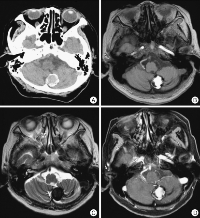
Fig. 1.
A : Computerized tomography scan shows a 2.5-cm in diameter, rim-calcified hemorrhagic lesion in the left cerebellar hemisphere. B : T1-weighted image reveals a 2.5-cm-sized well-defined hyperintense mass. C : T2-weighted image reveals a peripheral signal void rim and no perilesional edema. D : Gadolinium-enhanced T1-weighted enhancement image showing no contrast enhancement of the lesion.

