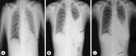
Fig. 1.
Serial chest X-ray. A : The shunt catheter is located in left upper quadrant of abdominal cavity and mild blunting of the left costophrenic angle is observed after initial ventriculoperitoneal shunt, B : The left pleural effusion with the suspicion of catheter migration into thoracic cavity is shown at second admission, C : After laparoscopic catheter reposition, the catheter is located in abdominal cavity and pleural effusion is lessened.

