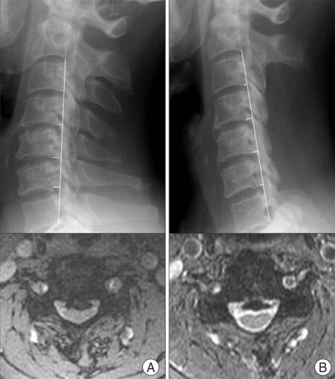
Fig. 2.
A-1, A-2 : Preoperative cervical spine X-ray and magnetic resonance (MR) image. B-1, B-2 : Postoperative cervical spine X-ray and MR image taken at the last follow-up examination. Pre- and postoperative curvature index values were calculated from lateral simple X-rays. The pre- and postoperative status of the cervical lesion and spinal cord were evaluated on MR images if there were other predisposing factors of neurological complications.

