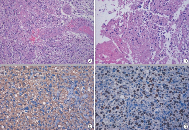
Fig. 2.
Histological examination of the removed tumor specimens showed a cellular tumor composed of elongated, spindle-shaped cells with irregular, moderately pleomorphic nuclei, as well as proliferative blood vessels and necrosis (A : original magnification ×100, B : original magnification ×200). The tumor cells showed cytoplasmic positivity for glial fibrilary acidic protein (C : original magnification ×200). Approximately 30-40% of the nuclei were positive for Ki-67 (D : original magnification ×200).

