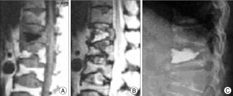
Fig. 1.
Imaging studies obtained from a 78-year-old woman with T12 compression fracture. A, B : Sagittal T1-and T2-weighted images showing the fracture cavity as discrete areas of abnormally low and high signal intensity, respectively. C : Lateral radiograph of the spine in a weight-bearing position obtained immediately after balloon kyphoplasty reveals effective filling of cystic fracture cavity.

