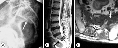
Fig. 2.
Postoperative cone view of the sacrum shows definite and aggravated fracture and displacement of the sacrum (A-arrow). Postoperative magnetic resonance imaging of the sacrum demonstrating aggravation of fracture and dislocation at the S1-S2 interspace (B-black arrow) and diffuse signal change at the sacral body and both sacral ala (C-white arrow).

