
Figure 3. Inhibitory effect of the p38 MAPK inhibitor SB203580 on HO-1 mRNA and protein induction after exposure of RASMCs to acidic media.
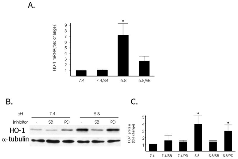
A. Quantitative analysis of HO-1 mRNA levels (normalized to 18s mRNA by Q-PCR) after exposure of RASMCs to acidic media (pH 6.8) compared to exposure to media of pH 7.4 with or without treatment with 5μM of SB203580. Cells were pretreated with 5μM SB203580 for one hour or DMSO (vehicle) and exposed to an extracellular pH of 6.8 or 7.4 for 8 hours. Data are expressed as the mean and standard error of the mean (SEM). Six independent experiments are represented. *p<0.05 compared to HO-1 mRNA at pH 7.4. B. Representative Western blot depicting HO-1 protein levels compared to levels of α-tubulin used as loading control. Cells were pretreated for one hour with DMSO (vehicle), the p38 inhibitor SB203580 (5μM-SB), or the Erk1/2 inhibitor PD098059 (10μM-PD) and were then exposed to an extracellular pH of 6.8 or 7.4 for 18 hours. C: Quantitative analysis of HO-1 protein levels after exposure of RASMCs to acidic media compared to exposure to media of pH 7.4 with or without treatment with 5μM of SB203580 or 10 μM of PD 098059. Data are expressed as the mean and standard error of the mean (SEM). *: p< 0.05, statistically significant difference as compared to pH 7.4, 7.4/SB, 7.4/PD and 6.8/SB. Six independent experiments are represented and quantitative analysis was done with NIH Image software and normalized to α-tubulin levels.