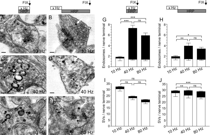
Figure 3.
HRP labels bulk endosomes when applied during strong action potential stimulation. A–F, HRP was applied to cultures during trains of 200 (A; 10 Hz), 400 (C; 40 Hz), or 800 (E; 80 Hz) action potentials and then immediately fixed. Alternatively, HRP was applied to cultures for 5 min immediately after trains of 200 (B; 10 Hz), 400 (D; 40 Hz), or 800 (F; 80 Hz) action potentials and then fixed. Representative electron micrographs are displayed. Black arrows indicate HRP-labeled endosomal structures, whereas white arrows indicate HRP-labeled SVs. Scale bars, 100 nm. Mean number ± SEM of either HRP-labeled (solid bars) or clear (open bars) endosomes (G) or SVs (I) generated during stimulation is displayed (200 action potentials, n = 17 nerve terminals; 400, n = 26; 800, n = 53). Mean number ± SEM of either HRP-labeled (solid bars) or clear (open bars) endosomes (H) or SVs (J) generated after stimulation is displayed per nerve terminal (200 action potentials, n = 18 nerve terminals; 400, n = 39; 800, n = 38). *p < 0.05, **p < 0.01, ***p < 0.001, one-way ANOVA for HRP structures.

