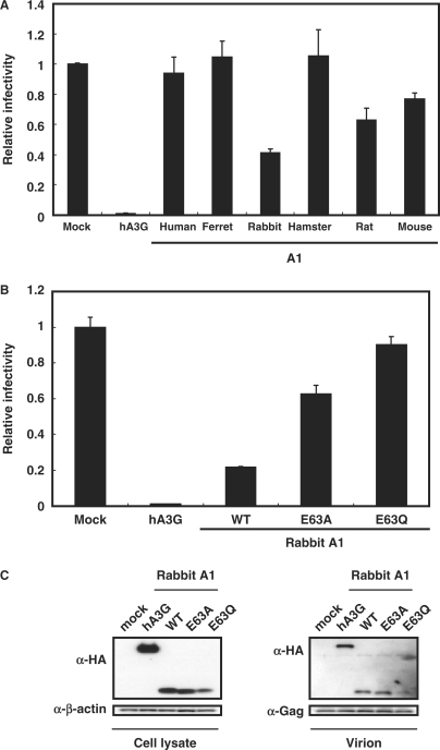
Figure 6.
Inhibition of MLV infection by A1s. (A and B) MLV packaging cell line GP293 were transfected with 1.5 μg of luciferase pFB-Luc reporter plasmids, 1.0 μg of pVSV-G and 0.5 μg of HA-tagged APOBECs or rabbit A1 with catalytic site mutations. Virus-containing supernatants were normalized for equal MLV p30 CA content and used for the infection of the MDTF cells. Virus-induced intracellular luciferase activity was measured and presented as described. (C) Rabbit A1 proteins are encapsidated into MLV virions. After transfection, released virion was collected by ultracentrifugation, while the producer cells were collected and lyzed. The cells and virion lysates were then subjected to Western analysis using antibodies specific for the HA tag and MLV Gag CA. An immunoblot probed with anti-β-actin antibody of the proteins present in the cell lysates is also shown. While only the immunoblot of p30 CA performed with the disrupted virions is presented, closely similar results were also obtained using the cell lysates (data not shown).

