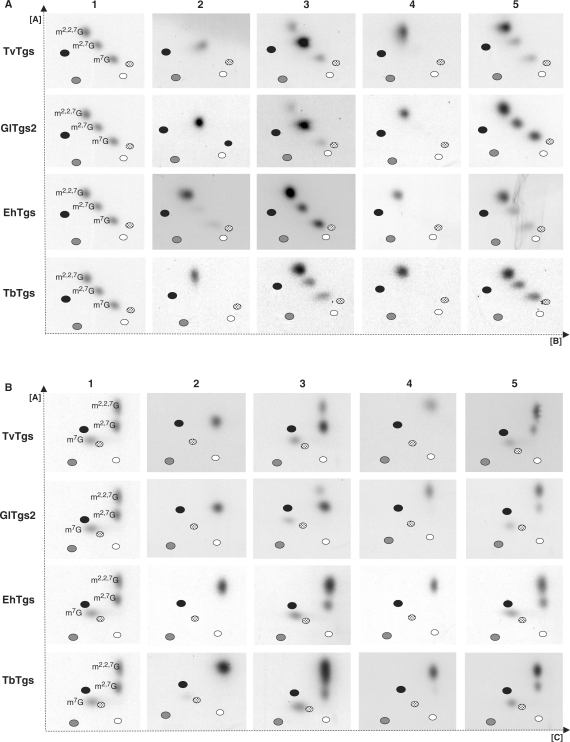
Figure 5.
2D-TLC analysis of cap structures formed using Tgs from four unicellular parasites. The first dimension was run using solvent A (28; see ‘Materials and methods’ section) and the second dimension used either solvent B or solvent C as shown in (A) or (B), respectively. 32P-labelled m7G cap RNA was the substrate for methyltransferase activity with recombinant Tgs from T. vaginalis (TvTgs), G. lamblia (GlTgs2), E. histolytica (EhTgs) and T. brucei (TbTgs) as indicated on the left. The guanosine capped end-products of these reactions were then immediately released by TAP treatment (columns 2 and 3) or were further incubated with S. pombe TgS (SpTgS) prior to release by TAP treatment (columns 4 and 5). Mono, di and trimethylguanosine standards were included as markers to reveal the relative migration of these cap structures (column 1). Black, grey, white and dotted ovals also indicate migration of the four monophosphate nucleotides—AMP, GMP, UMP and CMP, respectively. Samples were analysed in the absence (columns 2 and 4) or the presence of mono, di and trimethylguanosine standards (column 3 and 5) for clarity. In the latter case, the cap structure formed resulted in an increased intensity of the corresponding cap standard.

