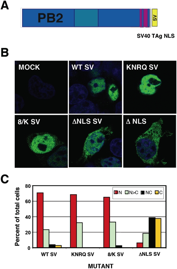
Figure 4. Intracellular localisation of mutated PB2 proteins containing an ectopic NLS.
(A) Diagram showing the structure of PB2 protein containing a SV40 TAg NLS at its C-terminus. (B) Representative phenotypes of the intracellular localisation of wt and mutant PB2 proteins. Cultures of HEK293T cells were transfected with plasmids expressing either wt or mutant PB2 proteins containing an additional NLS signal at the protein C-terminus. Central optical sections are presented of mock-transfected (MOCK), transfected with wt PB2 (WT) or with each of the mutant PB2 proteins. The phenotype of the ΔNLS mutant lacking the SV40 TAg NLS is also shown. Nuclei were stained with DAPI (blue) and PB2 was stained with anti-PB2 monoclonal antibody and goat anti-mouse IgG coupled with Alexa 488 (green) (C) Quantitative estimation of the localisation of wt or mutant PB2 proteins was performed as indicated in Fig. 2C.

