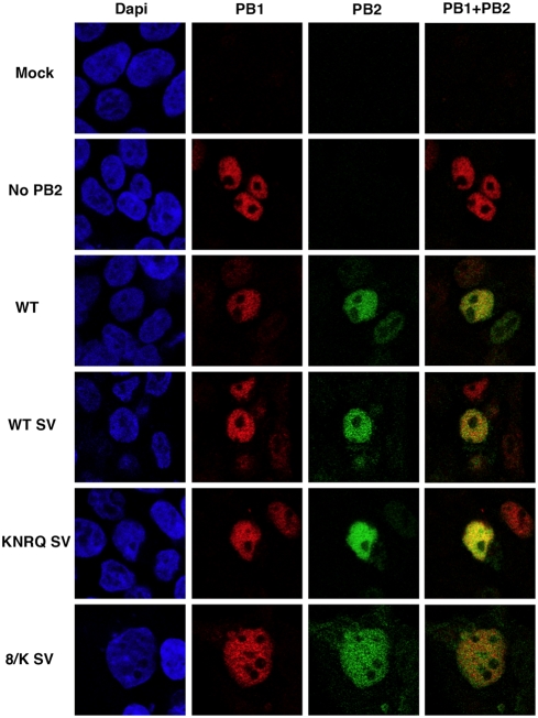
Figure 5. Intracellular localisation of wt and mutant PB2 proteins in cells co-expressing the polymerase subunits.
Cultures of HEK293T cells were co-transfected with plasmids expressing PB1, PA and either wt or mutant PB2 containing an additional TAg NLS. The cultures were fixed and analysed by double immunofluorescence using antibodies specific for PB1 and PB2 proteins. The signals for nuclear staining (DAPI –blue-), PB1 protein (PB1 –red-), PB2 protein (PB2 –green-) and the merge of PB1 and PB2 signals are shown for mock-transfected cells (Mock), cells expressing PB1 and PA (No PB2), cells expressing wt polymerase (WT), cells expressing polymerase with PB2-SV protein (WT SV) or cells expressing mutant polymerase containing each of the PB2 mutants with additional TAg NLS (KNRQ SV and 8/K SV).

