Figure 4.
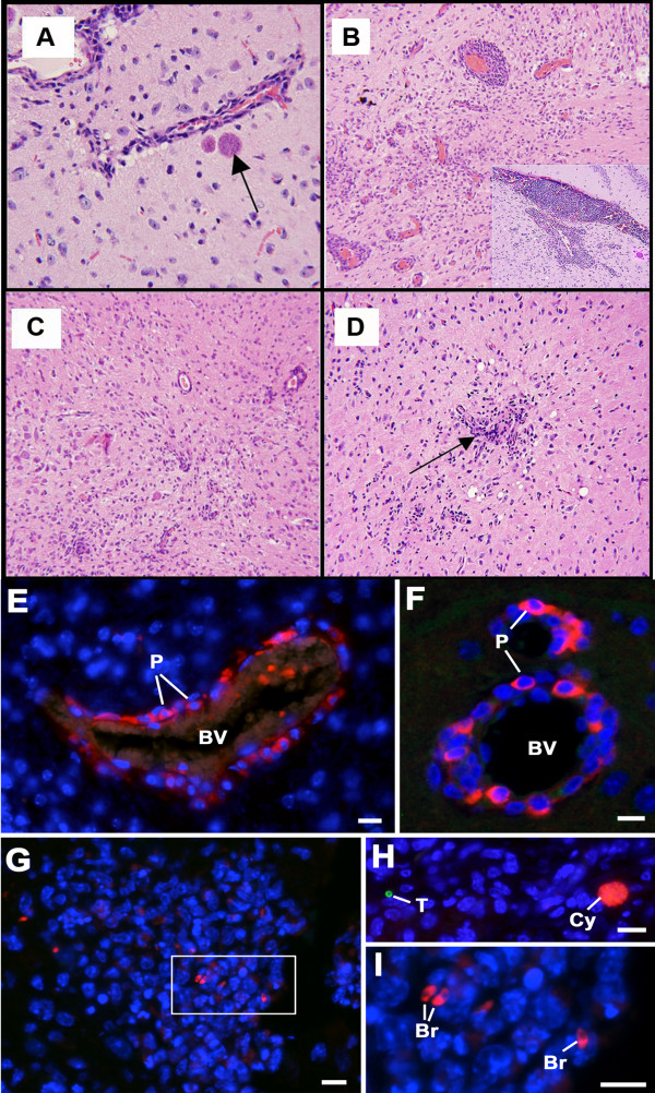
Inflammation in the brain during chronic Toxoplasma infection with only a few bradyzoites and cysts. (A) Occasional cyst with bradyzoites a short distance from a vessel (arrow), × 250. (B) Medium power view showing perivascular cuffing, × 100. (C) High power view of brain. Note perivascular cuffing and microglial infiltrates. Meningeal infiltrates also occurred (not shown), × 100. (D) Small numbers of microglial nodules (arrow) with diffuse inflammatory infiltrates throughout brain parenchyma, × 100. (E-I) Sections through the brains of chronically infected mice from the behavioral and neurologic studies, double labeled with rabbit anti-SAG1 visualised with anti-rabbit Ig conjugated to fluorescein isothiocyanate (green) and mouse anti-BAG1 visualised with anti-mouse Ig conjugated to Texas red (red). Bars represent 10 μm. (E, F) Examples of blood vessels (BV) cuffed with numerous inflammatory cells in which the plasma cells (P) can be identified by the cross reaction of the anti-mouse Ig with the immunoglobulins within the plasma cell cytoplasm (red), × 400. (G) Low power of a nodule of inflammatory cells in which a few bradyzoites (red) but no tachyzoites (green) could be identified, × 100. (H) A section of brain showing a single tachyzoite (green) and tissue cyst (red) in an area with no inflammatory cells, × 100. (I) Detail from the enclosed area in G showing the cytoplasmic staining of the bradyzoites (Br) with anti-BAG1, × 1000.

