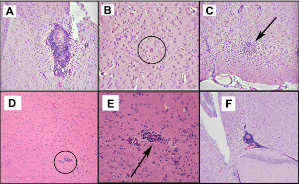
Figure 7.
(A-C) Similar histopathology with perivascular inflammation, isolated cyst ×40, and cluster of microglia in a mouse that is chronically infected, initially infected with parasites without accompanying brain. A ×40; B ×250; C ×20. (D) Less prominent perivascular cuffing (circle) and small collections of mononuclear cells in Balb/C mouse that is genetically more resistant. E. Increased magnification of area of perivascular inflammation. × 100.(F) Perivascular inflammation × 40 for mouse that had mild lateral ventricular dilatation in MRI.

