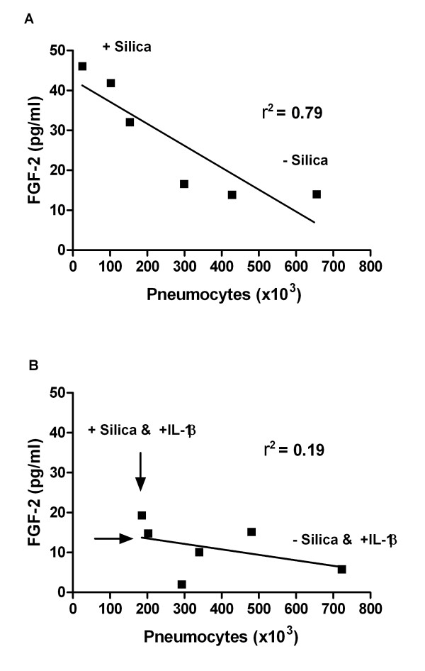
Figure 8.
The figure shows the correlation between changes in number of pneumocytes and FGF-2 release. These changes were measured in un-exposed and silica-exposed contact co-cultures (M+P) without (A) and with IL-1β supply (B). The IL-1β (75 pg/ml) was added to the contact co-cultures 30 min before silica exposure (160 μg/cm2). The time of exposure was 43 h. Data are from 3 separate experiments. A high r2 indicates a good correlation. The slope was only significantly different from zero in figure A (P = 0.0183). Arrows (B) indicate the directions of changes (e.g. increased cell number and decreased release of FGF-2). Univariate linear regression and correlation analysis were used to assess if cell number could explain the changes in release of FGF-2.

