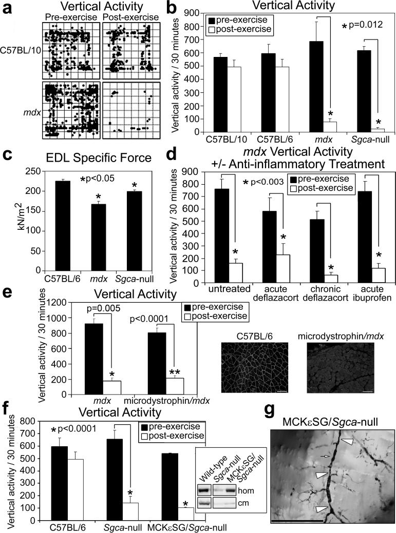
Figure 1. Loss of sarcolemma-localized nNOS leads to skeletal muscle vascular narrowings, reduced capillary perfusion, and an exaggerated fatigue response after mild exercise in dystrophic and non-dystrophic mouse models.
a, Representative C57BL/10 and mdx mice pre- and post-exercise zone map vertical activity tracings. b, Quantified vertical activity pre- and post-exercise for C57BL/10, C57BL/6, mdx and Sgca-null mouse strains (n=6 for each strain). c, EDL muscle specific force measurements from C57BL/6 (n=6), mdx (n=4), and Sgca-null (n=4) mice. d, Pre- and post-exercise vertical activity in untreated (n=7) and anti-inflammatory treated mdx mice, acutely (n=4) or chronically (n=4) with deflazcort or acutely with ibuprofen (n=5). Asterisks indicate statistical significance. e, Quantified pre and post-exercise vertical activity for Microdystrophin/mdx mice (n=6) and their mdx littermates (n=4). Insets show representative immunofluorescence images of nNOS detection in the gastrocnemius muscles from C57BL/6 and microdystrophin/mdx mice. f, Quantified pre and post-exercise vertical activity for MCKεSG/Sgca-null mice (n=6) and their Sgca-null littermates (n=6). Inset shows immunoblot detection of total nNOS from homogenates (hom), and crude skeletal muscle membranes (cm). g, Representative Microfil® image of skeletal muscle vessels of MCKεSG/Sgca-null mice post-exercise – large arrow heads mark extended areas of vascular narrowing and small arrows mark shorter stretches of radial vascular narrowing.

