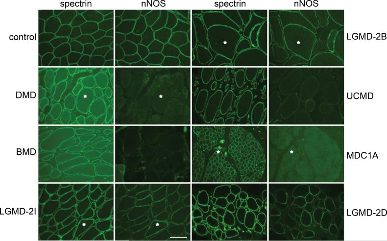
Figure 4. nNOS is reduced in human muscle diseases.
Representative immunofluorescent staining in various human muscle diseases: in primary dystrophinopathies, Duchenne and Becker muscular dystrophy (DMD and BMD, respectively); in several forms of limb-girdle muscular dystrophy (LGMD); in two congenital muscular dystrophies (CMD) caused by mutations in extracellular matrix proteins [Ullrich CMD (UCMD), collagen VI and merosin-deficient CMD (MDC1A), laminin-2]. Asterisks mark the same muscle fibers in some of the adjacent panels. (Scale bar = 100 μm)

