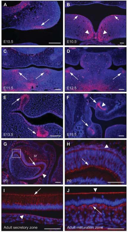
Figure 2.

Connexin 43 expression in the developing tooth germs of E10.5 through adult wild-type mice. (A,B) At E10.5, connexin 43 expression was observed in specific regions of the ventral epithelium of the developing maxilla (A; arrowed), in distinct regions of the dorsal epithelium of the developing mandible (B; arrowed), and also in the mesenchyme around the midline of the fusing mandibular processes (B; arrowheads). (C) At E11.5, connexin 43 expression was observed in the odontogenic mesenchyme of the mandibular processes (arrowed). (D) At E12.5, connexin 43 expression persisted in the condensing mesenchyme adjacent to the invaginating odontogenic epithelium (arrowed). (E) At E13.5, connexin 43 expression was detected in the lateral aspect of the bud-stage molar tooth germs (arrowed). (F) By E15.5, connexin 43 expression was observed in the lateral epithelium (arrowed) and in the enamel organ (arrowhead). (G,H) At P0, strong connexin 43 expression was apparent in the secretory ameloblasts (arrowed) and also in the stratum intermedium (arrowhead); weaker expression of connexion 43 was also detected in the stellate reticulum (sr). The boxed region in G is shown at a higher magnification in H. (I) In adult incisors, the secretory ameloblasts demonstrated strong, punctate expression of connexin 43 (arrowed); weaker expression was also detected in the stratum intermedium (arrowhead). (J) In the maturation zone of adult incisors, connexin 43 expression is restricted to the distal junctional complex of the ameloblasts (arrowhead), with strong expression observed in the stratum intermedium (arrowed). Scale bars in A-G = 100 μm; scale bars in H-J = 50 μm.
