
Fig. 8.
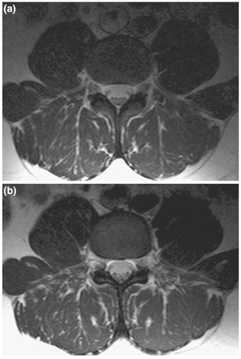
a T2-weighted lumbar spine axial image of a normal volunteer collected with the phi-coil: turbo-spin echo sequence with TE=90 ms, TR=2,550 ms, FOV=20 cm, matrix size = 256 × 196, slice thickness=5 mm, NEX=4. Signals from each detector element are combined by the sum-of-squares method. b The same anatomical region imaged with the figure-8 plus circular loop with identical imaging parameters and signal combination method