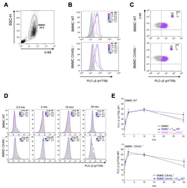
Figure 3. Tregs do not affect FcεRI-dependent PLC-γ phosphorylation.
WT or OX40L−/− sensitized BMMCs were stimulated with Ag in the presence of WT or OX40−/− Tregs for the indicated times. Cells were immediately fixed and stained for c-kit and phosphorylated PLC-γ2. From c-kit+-gated BMMCs (A), histogram overlays of phosphorylated PLC-γ2 at different time points were obtained from WT (upper panels) or OX40L−/− (lower panels) BMMCs challenged in absence of Tregs (B). Dot plot overlay of basal phosphorylated PLC-γ2 (left, gray) and after 10 min (right, violet) is shown in panel C. Histogram overlays of phosphorylated PLC-γ2 from WT (upper panels) or OX40L−/− (lower panels) BMMCs challenged in the presence of Tregs (D). Results shown are representative of three independent experiments. Kinetics of PLC-γ2 phosphorylation at different conditions are shown in panel E and are the mean + SD of three independent experiments.

