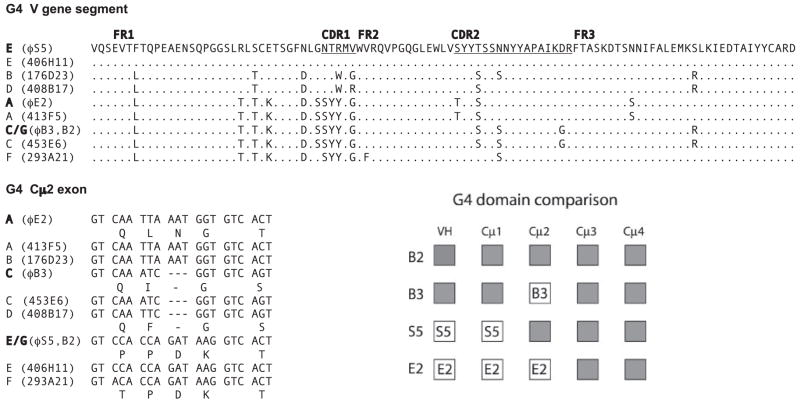
FIGURE 4.
Classification of G4 members by VH and Cμ2 sequences. Panel G4 V gene segment, The deduced amino acid sequences of VH sequences obtained from bacteriophage clones ϕE2, ϕB3/B2, and ϕS5 are compared among representatives of different BAC clones classified as G4A to G4F with their grid names. Dots indicate identity to reference VH sequence from ϕS5. Panel G4 Cμ2 exon, The 5′ end of the C2 exons of G4 genes are compared, and positions with different amino acids are indicated in the one-letter code. Dashes indicate gaps. Panel G4 domain comparison, The VH gene segments (VH, D1, D2, JH) are represented as a block, like the C exons. The domains of clones B3, E2, and S5 are compared with B2 to show the extent of shared sequence, indicated by shaded boxes. The domains of B2 and B3 are identical in all except Cμ2. E2 is identical to B2 in Cμ3 and Cμ4; S5 is identical to B2 in Cμ2, Cμ3, and Cμ4.

