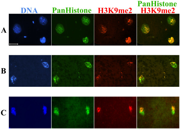
Figure 9.
H3K9me2 distribution in mouse (A), bovine (B) and rabbit (C) early two-cell embryos. H3K9me2 was visualized using a specific rabbit polyclonal antibody. Core histones were visualized by a mouse monoclonal anti-Pan histone antibody. DNA was counterstained with DAPI. In all presented cases H3K9me2 compartmentalized asymmetrically in the nuclei of early two-cell embryos (n > 5). Scale bar 20 μm.

