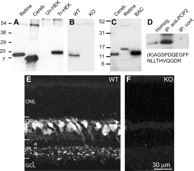
Figure 2.
The predicted new isoform of PCP2 is expressed in retinal ON bipolar cells. A, Western blots of proteins of retinal, cerebellar (cereb), and HEK cells. HEK cells were either untransfected (Un-HEK) or transfected with pcDNA3.1 Ret-PCP2 construct (Tr-HEK). The blot was probed with the antibody we generated against the N terminus region of exon2. PCP2 in extracts from retina and transfected HEK cells migrated with similar mobility, whereas a single band from cerebellum migrated faster. B, Western blots of WT and PCP-null (KO) retinas showed that the 15 kDa PCP2 band was eliminated in the null retina. C, Western blot containing protein lysates from retina and cerebellum as in A probed with an antibody against exon 3, a region common to all PCP2 isoforms. Lysate from cells expressing the large cerebellar form (1B) was included as a control (BAC). Note, only a single band appears in cerebellum and retina. D, Ret-PCP2 was immunoprecipitated with the antibody against the N terminus region of exon 2, isolated by SDS-PAGE and subjected to mass spectrometry (MS/MS) microsequencing analysis. This revealed a Ret-PCP2 unique peptide (shown below the blot) with a score xC = 2.54 (cutoff is 2.5). E, In the wild-type retina, staining for PCP2 localizes to somas high in the INL, whose axons terminate throughout sublamina b of the IPL. Staining in ON bipolar dendrites and axon terminals seemed equally strong. ONL, Outer nuclear layer; OPL, outer plexiform layer; INL, inner nuclear layer; IPL, inner plexiform layer; GCL, ganglion cell layer. F, In null retina, staining for PCP2 was totally eliminated.

