
Figure 2.
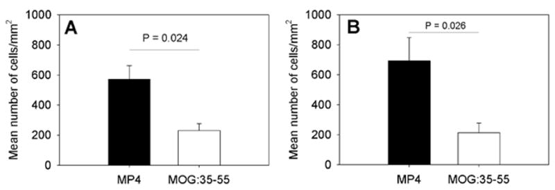
The density of cellular infiltrates in the CNS of MP4- and MOGp-immunized mice in acute and chronic EAE. WT B6 mice were immunized with MP4 (solid bars) or MOGp (open bars) as above. The CNS tissue was removed on the first day on which mice showed clinical symptoms of the disease and DAPI staining was performed on 7 μm thick cryostat sections. The mean total number of cells/mm2 ± SD infiltrating the CNS is given in panel (A) for acute EAE and for chronic EAE in panel (B). Results are shown for a total of 15 mice and 216 sections in each model and for each time point.