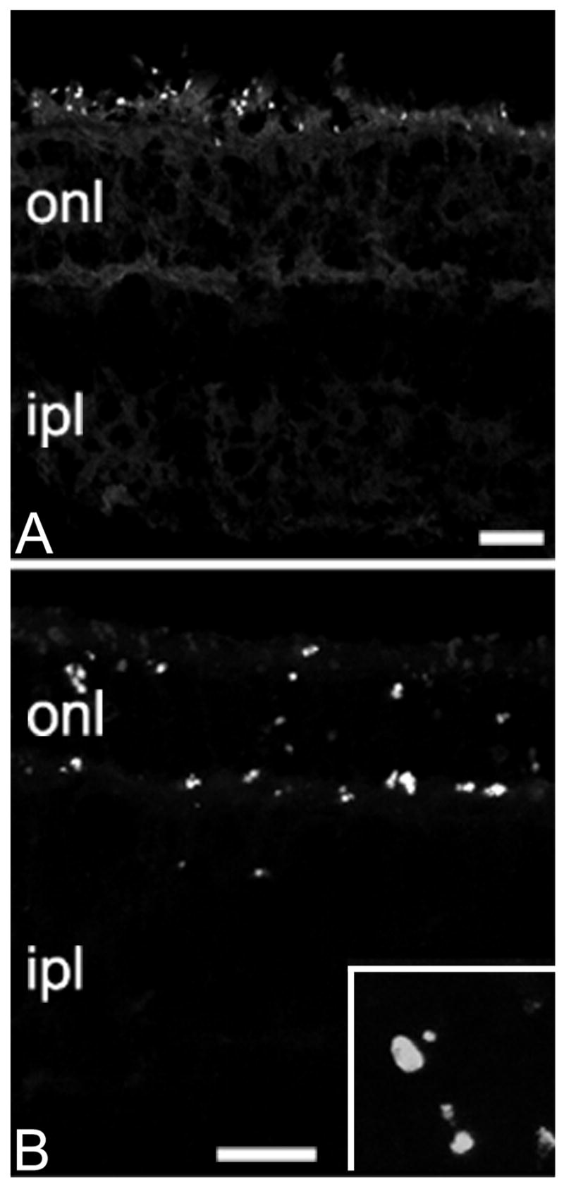
Figure 2.

Apoptotic markers in the photoreceptor layer. A: Fluorojade-positive profiles are scattered in the outermost portion of the retina at P20. B: photoreceptors stain positively with the TUNEL method at P25. Apoptotic profiles are clearly visible (inset: 4x enlargement of the onl, similar to that shown in B). Bars are 20 micrometers.
