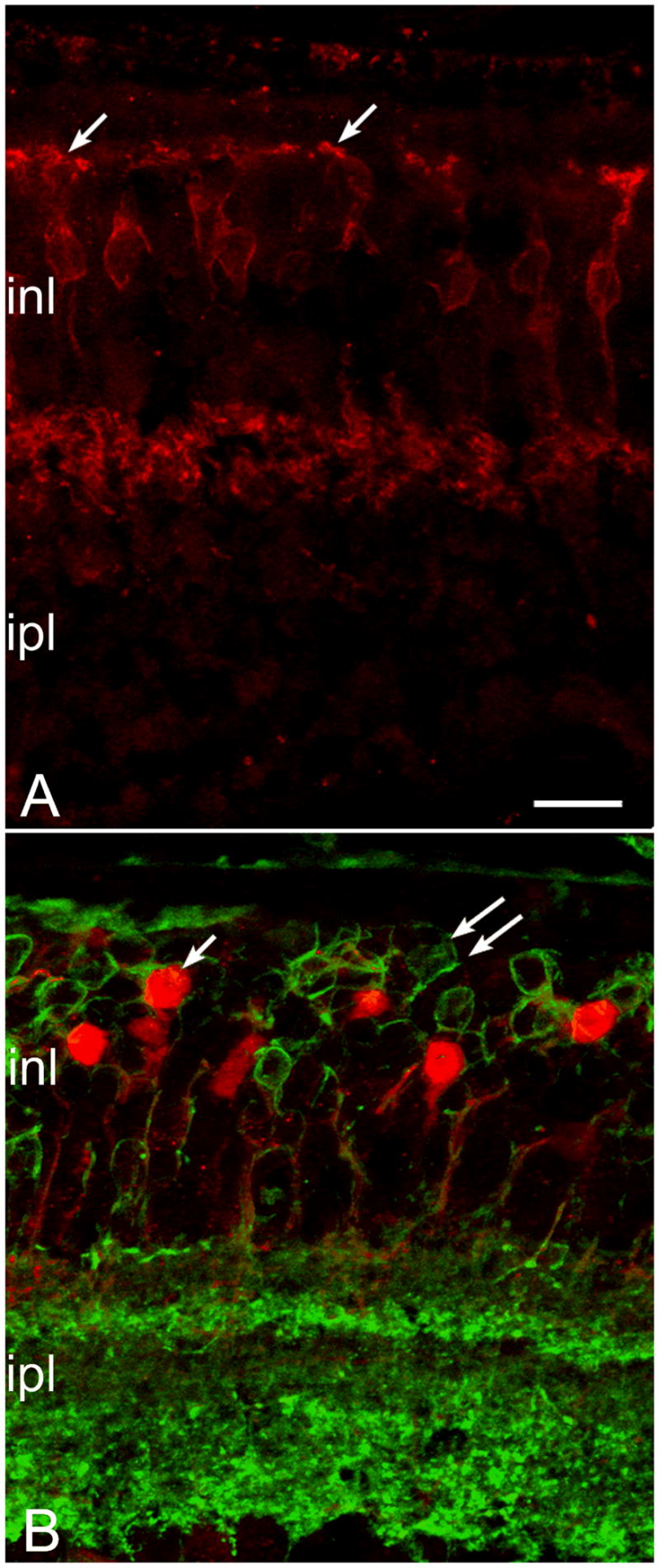
Figure 5.

Dendritic loss from cone bipolar cells at late stages of the rd10 degeneration. A: NK3-R staining of cone bipolar cells with axonal arbors in the outer half of the ipl shows normal dendritic morphology. B: In animals 9 months and older, cone bipolar cells of the same type, labeled with recoverin antibodies (red) have totally lost the dendrites; cell bodies appear completely smooth (left arrow). Green signal: Goalpha labeling of bipolar neurons show virtual absence of processes in the opl (double arrows). Bar is 20 micrometers.
