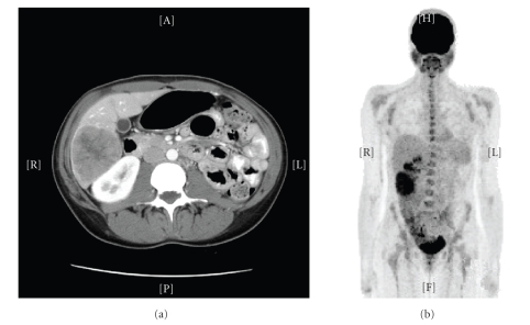
Figure 1.
CT and PET scans of EBV-related SMN of liver. CT scan of the abdomen and pelvis (8/05) from Patient 3 revealed multiple masses, mainly in the right lobe and involving segment 4. The largest mass measured 6.8 × 5.8 cm and was located in the right inferior lobe (a). FDG-PET (9/05) demonstrated areas of hypermetabolism in the inferior aspect of the right hepatic lobe (SUV max 5.7), and two other neighboring foci (SUV max 5) (b).

