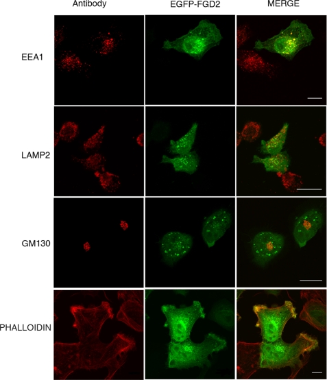
FIGURE 4.
Colocalization of FGD2 with organelle markers reveals partial localization to early endosomes and membrane ruffles. Shown are confocal fluorescent microscopy images of HeLa cells transfected with EGFP-FGD2 prior to fixation, permeabilization, and staining with antibodies against the endosomal marker EEA1 (upper panels), the lysosomal marker LAMP2 (middle panels), the cis-Golgi marker GM130 (lower panels) and phalloidin, a stain for F-actin (bottom panels). Bound antibody was detected with anti-mouse IgG Alexa 568 (red). A 10-μm scale is shown as white bars.

