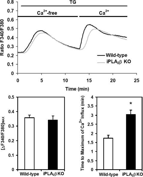
FIGURE 4.
TG-induced changes in intracellular [Ca2+] of wild-type and iPLA2β-null mouse aortic SMCs incubated in Ca2+-free or Ca2+-replete medium. First passage aortic SMCs from wild-type (WT) and iPLA2β–/– (KO) mice were grown to ∼70% confluency and then incubated in the dark at room temperature with 5 μm Fura-2/AM in a buffered solution (pH 7.3). The Fura-2/AM-containing buffer was then removed and replaced with Ca2+-free buffer containing 200 μm EGTA, and the cells were incubated for 10 min at room temperature to permit equilibration. Representative fluorescence tracings of intracellular [Ca2+] following addition of TG (1 μm) at t = 1 min in the presence of Ca2+-free buffer (t = 0–13 min) followed by continued incubation in the presence of fresh calcium- and TG-containing buffer added after 13 min. The magnitude of [Ca2+] entry was determined by ratiometric comparisons of the fluorescence intensities as a function of time after calcium re-addition. Intracellular [Ca2+] is expressed as the ratio of the fluorescence emission intensities at 510 nm achieved at excitation wavelengths of 340 nm and 380 nm (F340/F380) as described under “Experimental Procedures.” The maximal amplitudes of calcium entry (lower left panel) and the mean times required to achieve maximal Ca2+ amplitude (lower right panel) after adding Ca2+ to the medium of TG-treated cells are shown. Results presented are triplicate determinations obtained from each of three separate animals in each group and are expressed as the means ± S.E. *, p < 0.01.

