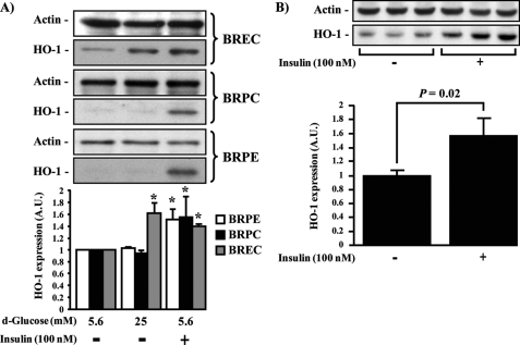
FIGURE 1.
HO-1 expression in retinal vascular cells induced by insulin. A, BREC, BRPC, and BRPE were treated with low glucose (5.6 mm), HG (25 mm) for 24 h, insulin (100 nm) for 6 h. B, intravitreous injection of saline (right eye) or insulin (10 μl of 100 nm; left eye) for 24 h in Sprague-Dawley rat retina (n = 6). Expression of HO-1 was detected by Western blot analyses and normalized with actin expression. One experiment representative of three immunoblots is shown with densitometric quantitation (means ± S.D.) from three independent experiments. *, p < 0.05 versus PBS-treated cells.

