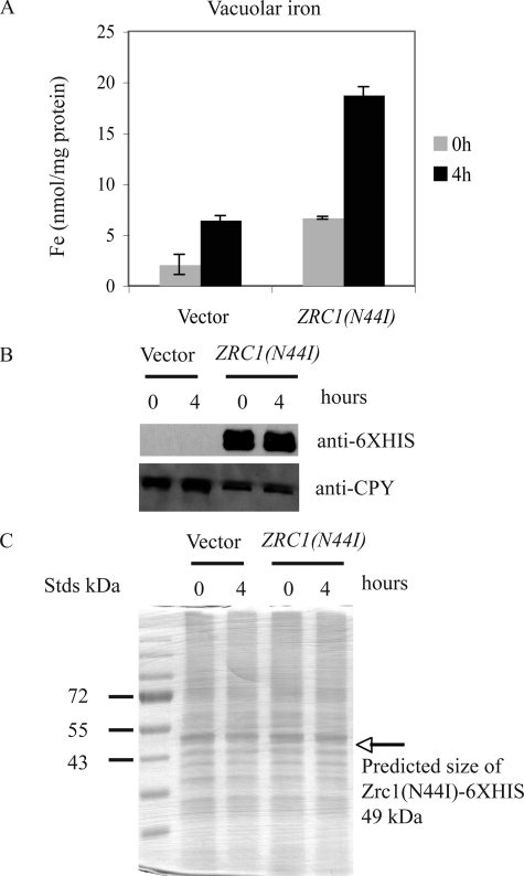
FIGURE 6.
Measurement of vacuolar iron content and Zrc1(N44I) protein abundance. A, Δccc1 cells were transformed with a control vector (pYES2) or a ZRC1(N44I)-HIS6 containing plasmid. Cells were grown in galactose CM overnight and then incubated in the same medium containing 100 μm FeSO4. Vacuoles were isolated from control and ZRC1(N44I) expressing cells at time 0 and 4 h after incubation in iron-rich medium. The iron content of isolated vacuoles was determined by ICP and the data normalized to protein content. B, isolated vacuoles were solubilized and analyzed by SDS-PAGE and Western blot using a rabbit anti-His6 antibody or a mouse anti-carboxypeptidase Y antibody followed by peroxidase-conjugated goat anti-rabbit IgG or goat anti-mouse IgG. C, the same samples as in B were also analyzed by silver staining. The arrow represents the predicted mass of Zrc1(N44I)-HIS6.

