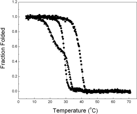
FIGURE 2.
Temperature melt curves of the collagen fragments. The thermal unfolding of F877 (square), G901S (circle), and G913S (triangle) was monitored by the temperature-induced decrease of the CD signal at 225 nm, and normalized to the fraction folded of the triple helix. The heating rate is ∼0.1 °C/min, and the concentrations of the three fragments are 1 mg/ml.

