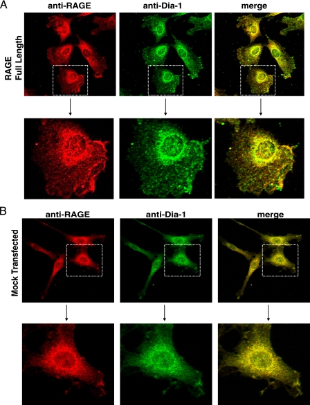
FIGURE 3.
RAGE and Dia-1 colocalization in cells. Confocal microscopy was performed using the C6 cells stably transfected with either full-length RAGE (A) or empty vector (Mock) (B) constructs. Cells were treated with RAGE ligand for 30 min, fixed, and immunostained with antibodies to RAGE and Dia-1 followed by respective biotinylated secondary antibodies and streptavidin-linked Alexa-546 (anti-RAGE) and 488 (anti-Dia-1). Higher magnification images of the cells within the stippled boxes in the upper panels are illustrated in the lower panels.

