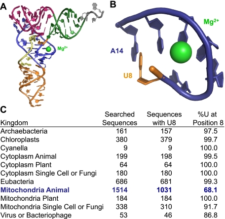
FIGURE 10.
The location of the Mg2+ binding pocket in tRNAPhe involving the highly conserved U8. A, tertiary structure of yeast tRNAPhe showing the Mg2+ (green) bound near U8 (orange). The tRNA is colored by secondary structure domains. The acceptor arm is green; the T-arm is pink; the D-arm is blue; the anticodon arm is orange; the variable loop is yellow; and the ACCA end is gray. B, close-up view of the Mg2+ binding pocket colored as in A. C, conservation of U8 among the tRNAs from various organisms and organelles. The conservation of U8 in mammalian mitochondrial tRNA is shown in blue.

