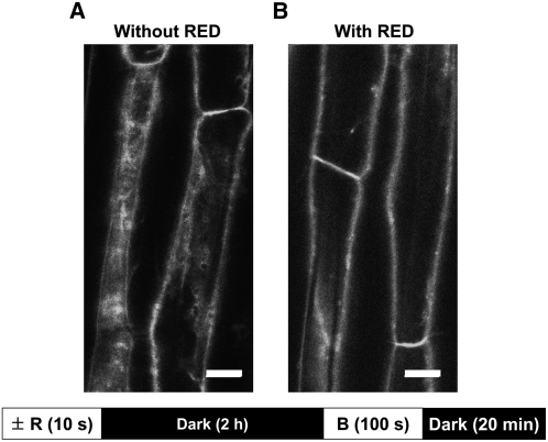
Figure 4.
Effect of Red Light Treatment 2 h before Blue Light Irradiation on Blue Light–Induced PHOT1-GFP Relocalization in 3-d-Old Arabidopsis Hypocotyl Cortical Cells.
Single z-series images taken from seedlings without (A) or with (B) red light (R) treatment 2 h before the blue light (B) pulse. Red light source as in Figure 1B. The blue light source (fluence rate 20 μmol m−2 s−1, exposure time 100 s) was LEDs. Single z-series optical sections obtained 20 min after the blue light pulse. Note minimal cytoplasmic PHOT1-GFP in red light–treated sample. Bar = 20 μm.

