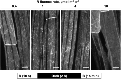
Figure 6.
Fluence Response Relationship for Red Light–Mediated Inhibition of Blue Light–Induced PHOT1-GFP Relocalization.
Exposure time constant (10 s), fluence rate varied (from 0.4 to 10 μmol m−2 s−1). Projection images of PHOT1-GFP in hypocotyl cortical cells from 3-d-old etiolated Arabidopsis seedlings. Note that 100 μ m−2 (10 s × 10 μmol m−2 s−1) of exposure to red light is sufficient to prevent blue light–induced PHOT1-GFP relocalization. The blue light source was the laser from a confocal microscope. Bars = 20 μm.

