
Figure 7. miR-132 modulates Per1 gene transcription and Per2 protein stability.
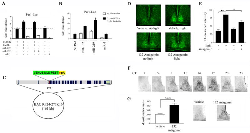
(A) Effect of miR-132 and miR-219 on CLOCK/BMAL1-mediated Per1 transactivation. HEK293T cells were transfected with a Per1-luciferase reporter construct, in combination with the following: CLOCK and BMAL1 expression plasmids and constructs encoding pre-miR-132, pre-miR-219 or pre-miR-1. Cells were lysed 48 hrs post-transfection and assayed for luciferase activity. Y-axis denotes fold difference in luciferase activity relative to control samples transfected with the Per1-luciferase construct alone. Experiments were performed four times, and representative data were averaged from quadruplicate determinations. (B) Effect of miR-132 and miR-219 expression on Per1 transactivation in primary neurons under basal and stimulated conditions. Primary rat embryonic neurons were transfected with a Per1-luciferase reporter construct in combination with the expression vector for pre-miR-132, pre-miR-219 or pre-miR-1. Forty-eight hrs post-transfection, neurons were stimulated with 25 mM KCl and 5 μM forskolin and cell lysates were prepared 6 hrs later. Y-axis denotes fold difference in luciferase activity relative to control samples transfected with the Per1-luciferase construct alone. Experiments were performed four times, and representative data were averaged from quadruplicate determinations. (C) Generation of Per1-Venus BAC transgenic mice. The Venus-NLS-PEST-polyA cassette was inserted into the per1 locus on a 161-kb bacterial artificial chromosome (BAC) by homologous recombination. The BAC clone contains the full intergenic sequences of mPer1 and its flanking genes, including the 5′ promoter region. (D) The effect of miR-132 knockdown on light-induced Venus expression in the SCN of Per1-Venus BAC transgenic mice. miR-132 antagomir (40 μM, 3 μl) or drug vehicle (saline), were infused into the lateral ventricles of Per1-Venus BAC mice at CT 14, and a brief light pulse was administered at CT 15. Tissue was harvested at CT 19 for analysis of Venus expression by immunofluorescence. Dark control animals were not exposed to light but were sacrificed at the same circadian time. (E) Quantitation of light-induced Venus expression as a function of miR-132 abundance. Values are presented as mean ± SEM. n=3–8 mice per group. *p<0.05 (two-tailed Student’s t-test). (F) Immunohistochemical analysis of PER2 protein abundance in the SCN as a function of circadian time. (G) In a separate experiment, mice were infused with miR-132 antagomir (40 μM, 3 μl) or vehicle at CT 14 and exposed to light (100 lux, 15 min) at CT 15 (right). SCN tissue was harvested 29 hr later and immunostained for PER2. Quantitation of PER2 immunoreactivity in the SCN of miR-132 antagomir- or vehicle-infused mice (left). Values are presented as mean ± SEM. n=8–10 per group. *p<0.05 (two-tailed Student’s t-test).