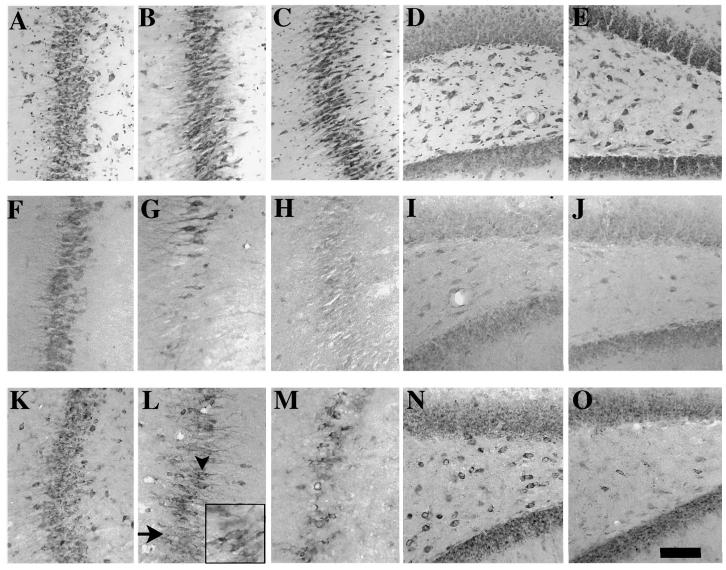
FIG. 3.
Bcl-2 and Bax immunoreactivities in the hippocampus following lateral fluid-percussion brain injury. Photomicrographs from representative serial sections from the CA3 region of sham-injured animals (A,F,K) and brain-injured animals at 2 h (B,G,L) and 6 h (C,H,M) post-injury, stained with Toluidine Blue (A–C), and anti-Bcl-2 (F–H) and anti-Bax (K–M) antibodies. Photomicrographs from representative sections illustrating the hilus of the dentate gyrus in sham-injured animals (D,I,N) and at 2 h post-injury (E,J,O) stained with Toluidine Blue (D,E), and, anti-Bcl-2 (I,J) and anti-Bax (N,O) antibodies. At 2 h (B) and 6 h (C) after brain injury, neurons in the CA3 region take on a shrunken elongated shape suggestive of damage. While a significant number of cells in this region lose Bcl-2 immunoreactivity at 2 h (G) and 6 h (H), only a few cells lose Bax immunoreacivity at 2 h (L, arrow). Bax(+) cells in the CA3 region at 2 h post-injury (L, arrowhead) stain in a pattern similar to that in sham animals, with diffuse staining in the cytoplasm (L, inset). At 6 h post-injury, however, the number of cells that do not stain for Bax protein have increased (M). Bar = 100 μm (all panels) 170 μm (inset in L).

