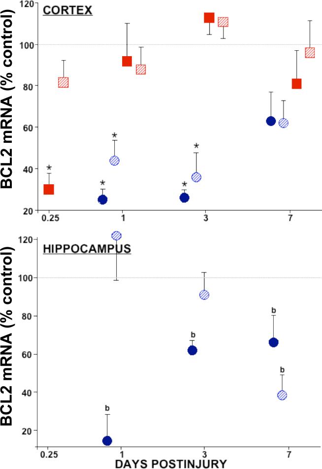
Figure 1. Relative levels of bcl-2 mRNA in cortex and hippocampus after traumatic brain injury.
In the injured cortex (top), bcl-2 mRNA decreased 70−80% at 0.25 d after LCI (solid squares) and from 1 to 3 d after LFP (solid circles) TBI. Contralateral cortex showed no changes (LCI, striped squares), or 50−60% decreases in bcl-2 mRNA from 1 to 3 d postinjury (LFP, striped circles). Hippocampal bcl-2 levels (bottom) were constitutively below (LCI, not shown) or dropped below the limit of quantitation (b) after LFP TBI. Relative mRNA levels (mean ± SEM) are the mole percent of cyclophilin mRNA, normalized to sham controls. Ipsilateral: solids, Contralateral: stripes. *p<0.05 vs. shams, ANOVA, Dunnett.

