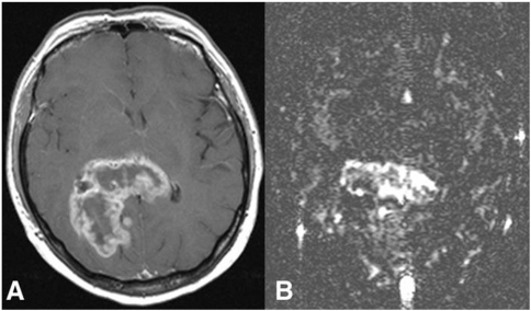
Figure 3.
(A) Transverse contrast-enhanced T1-weighted MR image shows a rim-enhancing tumour, histologically glioblastoma multiforme. (B) Blood flow map obtained with arterial spin labelling reveals high heterogeneity in total blood flow, which is seen highest in the central portion; from Warmuth et al.[80] with permission from the Radiological Society of North America.

