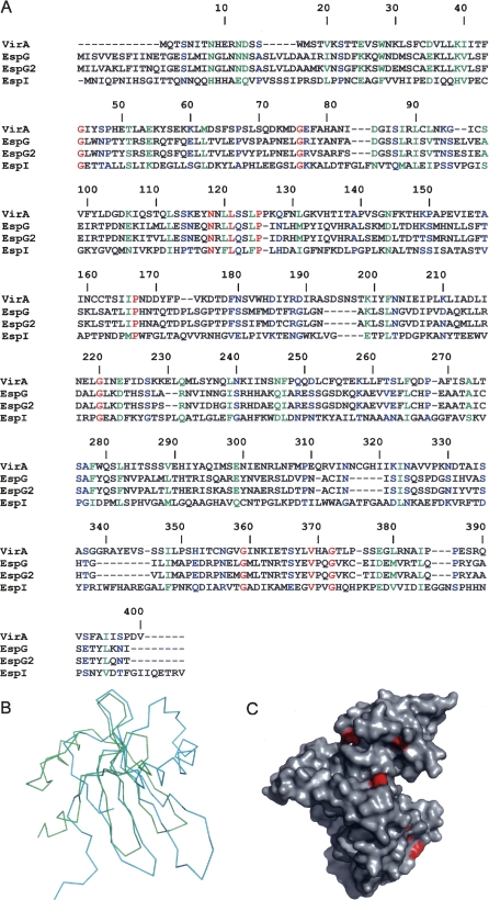
Figure 2.
Conservation of the sequence in the VirA/EspG family. (A) Sequence alignment of VirA, EspG, EspG2, and EspI proteins performed using ClustalW. Red denotes identical, green denotes strongly similar, and blue denotes weakly similar conservation. (B) Superposition of the Cα coordinates of the N-terminal domain of VirA and the inhibitor stefin B from the complex with papain (PDB code 1STF). VirA is traced in green; stefin B, in cyan. (C) Surface diagram of VirA in which the residues that are strictly conserved among VirA, EspG, EspG2, and EspI are colored red.

