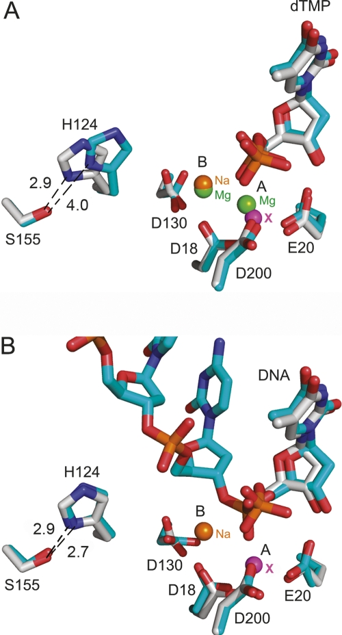
Figure 4.
Histidine 124 in the inhibited and active complexes including the structure with single-stranded DNA. Active-site superimposition of the inhibited sodium complex (gray) with (A) the catalytic magnesium complex (blue) and (B) the TREX1–ssDNA complex (blue). The protein residues and the dTMP molecule are labeled and represented as atom-type sticks. The metal-binding sites A and B are also indicated, and the spheres representing the ions are labeled in green (magnesium), orange (sodium), and magenta (undetermined metal). Distances between His124 and Ser155 are clearly indicated. Coordinates of TREX1–ssDNA were taken from the PDB code 2O4I.

