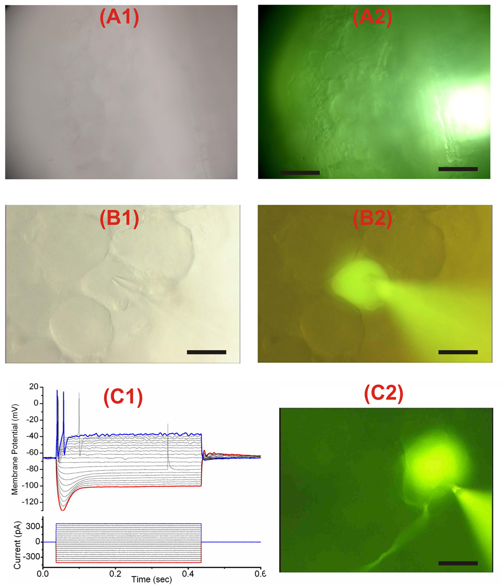
Figure 2. Visualization and recording from DRG neurons.
(A1–A2) Images of DRG neurons, visualized with 40X objective, were taken using transillumination (A1) and oblique epi-illumination (A2). This latter type of illumination enhanced the contrast and resolution of the image of neurons especially of thick DRGs where transmitted light was not effective for clear visualization. Epi-illumination was achieved by reducing the aperture diaphragm to a minimum and by slightly offsetting the light. It results in an increase in contrast and resolution, and the production of a shadowed, relief-like pseudo 3D appearance in the image of the DRG somata. Scale bar is 160 µm in (A1) and (A2). (B1) and (B2) DRG neuron visualized with trans-illumination which works adequately in this instance because the neuron was located superficially at the periphery of the ganglion. (C1) Membrane potential responses to 400 ms current injection of different amplitude from −400 pA to +400 pA in a DRG neuron filled with Lucifer Yellow shown in (C2). Scale bar is 20 µm in (B1), (B2) and (C2).

