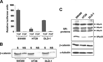
FIGURE 1.
Activation of β-catenin/TCF signaling in colorectal cells lines. (A) Diagram showing the activity of TOPglow, a β-catenin/TCF-regulated transcriptional luciferase reporter construct (TOP) or a mutant control construct (FOP) after transfection into the three indicated colorectal cell lines. Results were normalized to the activity of a co-transfected, constitutively active Renilla luciferase construct. (B) Western blot showing the comparison of the nuclear chromatin-associated β-catenin pool in the three cell lines. A detergent-based cell fractionation methodology separated β-catenin into a soluble (S) and a nonsoluble, chromatin-bound (NS) pool. Note that the amount of non-soluble nuclear β-catenin correlated well with the corresponding TOPglow activity in each cell line. (C) Western blot analysis of the pattern of SR protein expression in the three cell lines. The pan-SR protein antibody 1H4 was used to detect the indicated endogenous SR proteins. Levels of β-catenin and α-tubulin were detected as loading controls.

