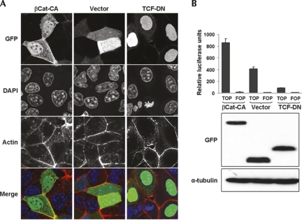
FIGURE 2.
Characterization of GFP-tagged β-catenin and TCF-4 mutants and their effect on β-catenin/TCF transcriptional activity. (A) Subcellular localization of the mutants by confocal microscopy. Images show DLD-1 colorectal cells transfected with (left column) a non-degradable, constitutively active β-catenin mutant (βCat-CA), (middle column) the empty pEGFP control vector (vector), or (right column) a dominant-negative TCF-4 mutant (TCF-DN). Note the nuclear localization of βCat-CA and TCF-DN. (B) Variation in the endogenous β-catenin/TCF transcriptional activity of DLD-1 cells. (Upper panel) A diagram of the TOPglow and FOPglow reporter activities upon co-transfection with βCat-CA, empty pEGFP vector, or TCF-DN. (Middle and lower panels) Western blot analyses of the GFP-βCat-CA, pEGFP, and GFP-TCF-DN expression levels as well as α-tubulin as loading control. Note the increased luciferase activity in the presence of GFP-βCat-CA and the inhibition by TCF-DN.

