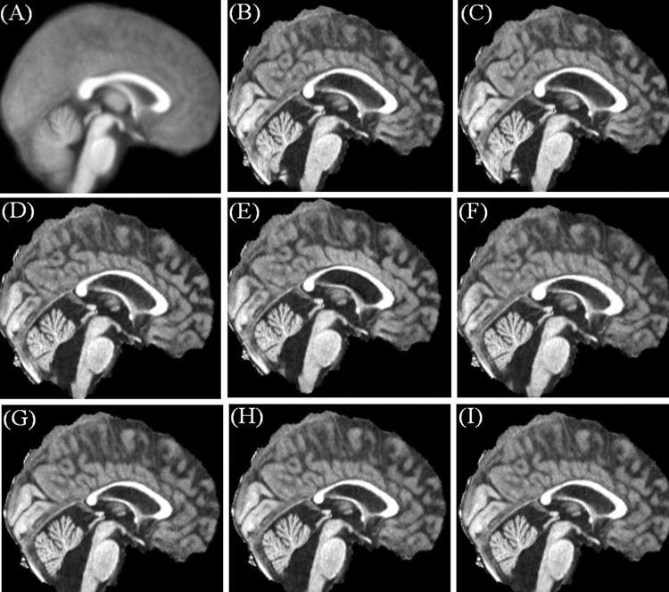
Fig. 2.
Comparison of the accuracy of the FFD spatial normalization approach with that of the DCT approach in a PFBT survivor with enlarged ventricles and the surgical void in the cerebellum. (A) Customized template. (B) Brain image after the affine transform using the FFD approach. (C) Brain image after the affine transform using the DCT approach. (D)–(F) FFD normalized brain images using the control point spacing distances of 20, 15 and 10 mm. (G)–(I) DCT normalized brain images using the cutoff frequencies of 1/20, 1/15 and 1/10 mm−1.

