Figure 1.
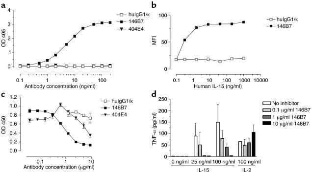
Recognition of receptor-bound IL-15 by mAb 146B7 and inhibition of IL-15–induced effects. (a) Biotinylated mAb 146B7 (filled squares) showed dose-dependent binding to IL-15 bound to IL-15Rα, which was coated onto an ELISA plate, whereas biotinylated 404E4 (filled triangles) did not show binding. Polyclonal huIgG1 (open squares) served as a control. This experiment was repeated two times, yielding similar results. (b) Dose-dependent binding of biotinylated mAb 146B7 (filled squares) to IL-15 bound to Raji lymphoma cells expressing IL-15Rα was shown by flow cytometry. Polyclonal huIgG1 (open squares) was used as a control. This experiment was repeated five times, yielding similar results. MFI, mean fluorescence intensity. (c) Effect of mAb 146B7 on IL-15–induced proliferation. Human PBMCs were incubated with IL-15 (12.5 ng/ml) in combination with mAb 146B7 (filled squares), mAb 404E4 (filled inverted triangles), or with huIgG1 (open squares) for 72 hours. BrdU incorporation was measured to assay proliferation. Representative data of 9 individual experiments are shown. (d) mAb 146B7 inhibits IL-15– but not IL-2–induced TNF-α production. Human PBMCs were incubated with IL-15 (0, 25, 100 ng/ml) or with IL-2 (100 ng/ml) in combination with mAb 146B7 at various concentrations for 72 hours. The amounts of TNF-α produced were measured by ELISA. A representative experiment from a series of 11 experiments is shown.

