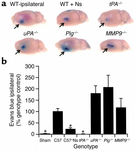
Figure 2.
BBB permeability following cerebral ischemia. (a) Analysis of Evans blue dye extravasation in mice. Brains were removed and photographed 6 hours after MCAO. All images are ipsilateral to the ischemic area. WT ipsilateral is a WT C57BL/6J mouse, and WT + Ns is a WT C57BL/6J mouse treated with an intraventricular injection of 2.5 μl of 16 μM neuroserpin immediately after MCAO. tPA–/–, uPA–/–, Plg–/–, and MMP9–/– are mice deficient in the enzyme indicated. The arrows indicate the area of Evans blue extravasation associated with the ischemic injury. (b) Quantitative analysis of Evans blue extravasation from brain extracts 6 hours after MCAO. Sham indicates animals subjected to all procedures except MCA ligation. The results represent the specific absorbance of Evans blue at 620 nm calculated as percentage of that of the WT control, either C57BL/6J (C57) or 129S6/SvEv as described in Methods. As a control for perfusion efficiency, the absorbance of the contralateral hemisphere was subtracted from that of the hemisphere ipsilateral to the MCAO. For each condition, n = 4. *P < 0.05 relative to WT control mice receiving MCAO.

