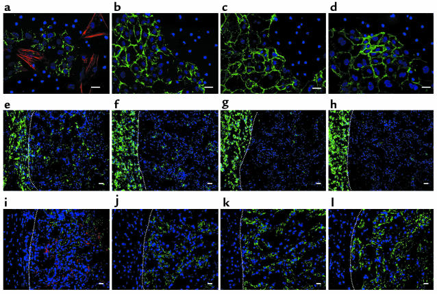
Figure 7.
Role of exogenous monocytes in EMT of renal tubular epithelial cells. (a–d) Dual immunofluorescence of E-cadherin (green) and α-SMA (red) in renal tubular epithelial cells and bone marrow monocytes cocultured for 48 hours. (a) WT epithelial cells and WT monocytes. (b) WT epithelial cells and Smad3-null (KO) monocytes. (c) KO epithelial cells and WT monocytes. (d) KO epithelial cells and KO monocytes. (e–l) Three days after transplantation of monocytes into the subcapsular space of the kidney with UUO. Dotted lines indicate the border between the subcapsular space (left) and the renal cortex (right). (e–h) Immunofluorescence of F4/80 antigen (green). (i–l) Dual immunofluorescence of E-cadherin (green) and α-SMA (red). (e and i) Transplantation of WT monocytes into WT kidneys. (f and j) Transplantation of KO monocytes into WT kidneys. (g and k) Transplantation of WT monocytes into KO kidneys. (h and l) Transplantation of KO monocytes into KO kidneys. DAPI (blue) was used for nuclear staining. Scale bars: 20 μm. Similar results were obtained from four additional experiments.

