Figure 2.
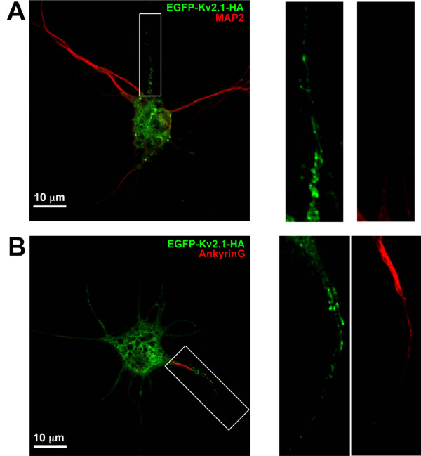
The single proximal neurite containing Kv2.1 corresponds to the axon initial segment. Hippocampal neurons grown for 10 days were transfected with EGFP-Kv2.1-HA and fixed 18 h later. Cultures were then labeled with either anti-MAP2 or anti-AnkG followed by Alexa 594-conjugated secondary antibodies. Shown in panel A is an example of Kv2.1 clustering in the proximal axon as indicated by the diminished MAP2 staining. Panel B illustrates intense AnkG staining in the Kv2.1 positive process. The right hand panels contain enlarged images of the neurites boxed in Panels A and B. Note that the diffuse GFP signal in the neurites represents intracellular GFP-Kv2.1.

